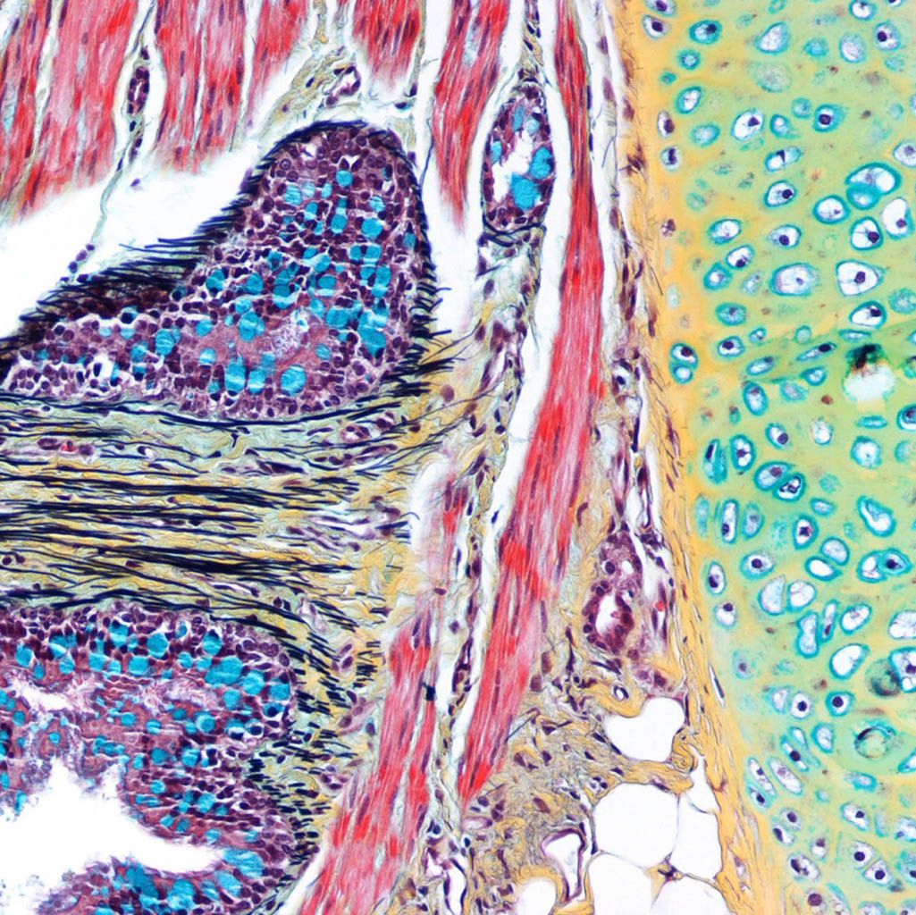
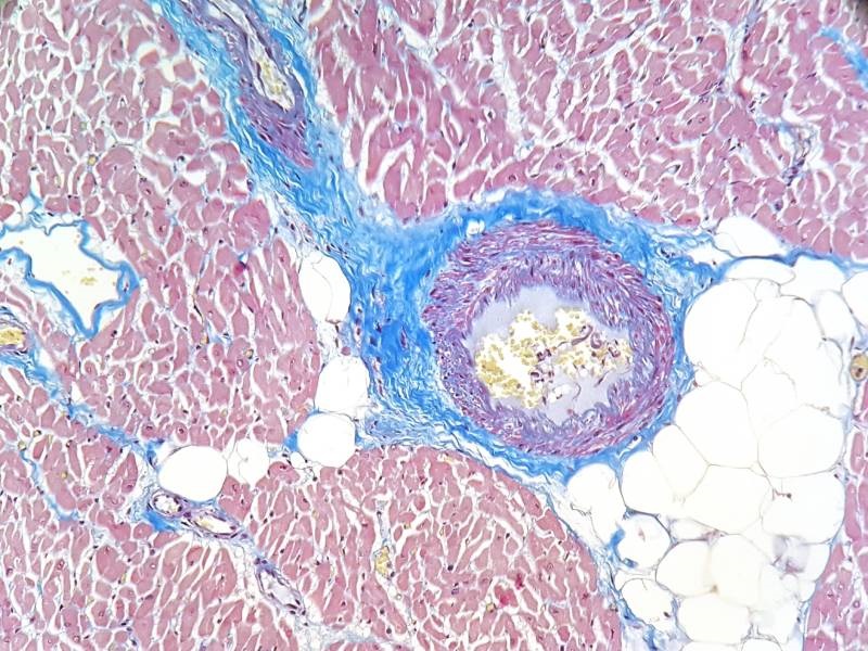
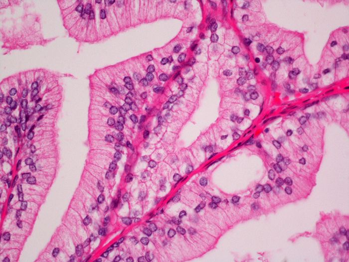
Sirius Red Stain Kit (Connective Tissue Stain) is intended for use in the histological visualization of collagen I and III fibers in addition to muscle in tissue sections.
The sirius red staining may be viewed using standard light microscopy.
Sirius red staining has traditionally sometimes been used with polarized light to more easily differentiate the collagen fibres from the background. Studies have used the differences between the appearance of different collagen fibres under polarized light to differentiate between different types of collagen fibres; it is unclear if the differences in appearance of the fibres is due to orientation, thickness and packing of the fibres rather than reflecting different types of collagen fibers.
Resources